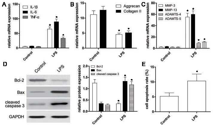
Fig. 2. LPS induces inflammatory cytokines, extracellular matrix degradation and apoptosis in NP cells. (A) qRT-PCR was used to detect the mRNA expressions of inflammatory cytokines (IL-1β, IL-6 and TNF-α) in LPS-treated NP cells; GAPDH was used as an internal control. (B,C) qRT-PCR was used to detect the mRNA expressions of ECM genes (Aggrecan, Collagen II) and matrix degrading enzymes (MMP-3, MMP-13, ADAMTS-4, and ADAMTS-5). (D) Western blot was used to detect the expressions of apoptosis-related proteins (Bcl-2, Bax and cleaved caspase 3) of NP cells. (E) Annexin/PI assay was used to measure the apoptosis rate of NP cells. Each experiment was assayed in triplicate independently. Values were presented as the means ± SD. *P<0.001, v.s. control.